


Researchers have developed a PET tracer that can visualize pathological alpha-synuclein aggregates in the brain. The method could contribute to the development of new therapies and help tailor personalized treatments in the future.
“With this new method, we can observe pathological processes directly in the brain for the first time – and further research will show whether this is also possible before clinical symptoms become clearly apparent,” says Kristina Herfert of the Werner Siemens Imaging Centre at Tübingen University Hospital, who led the study together with Armin Giese and MBExC member Christian Griesinger. “This opens up new possibilities for earlier and more accurate diagnosis.”
You can find the press release (in German) here.
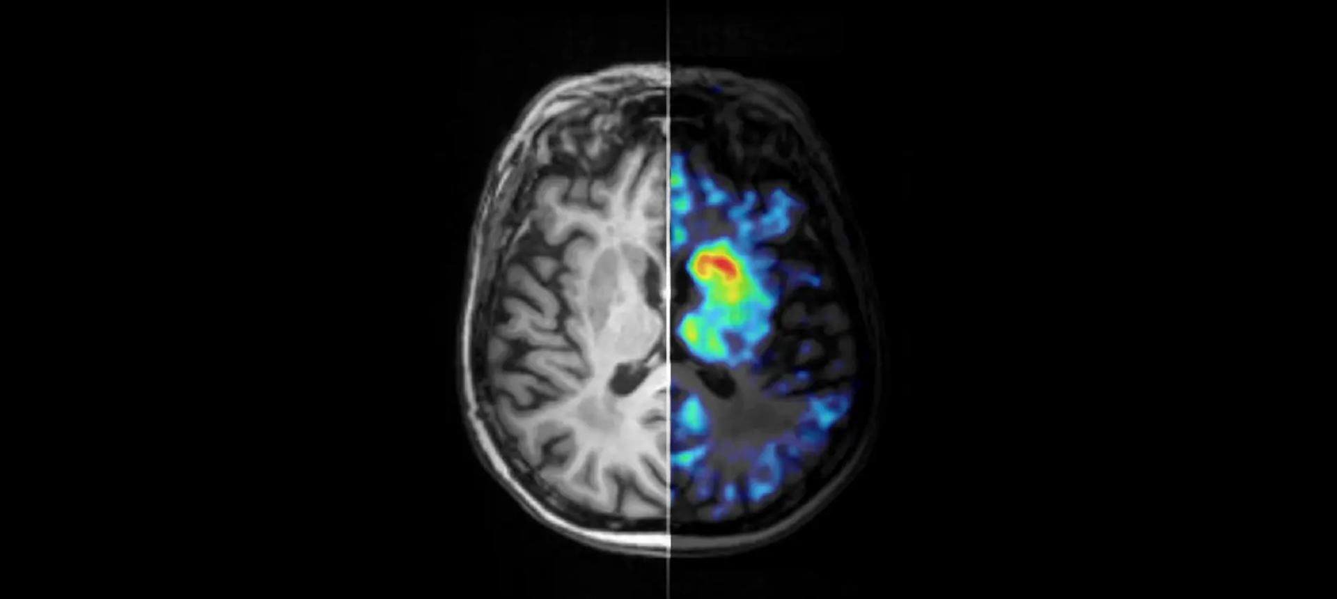
A novel alpha-synuclein PET tracer, [11C]MODAG-005, visualises pathological protein deposits in the human brain. The split-screen image compares the anatomical brain structure (left) with the molecular pathology (right) and illustrates the selective tracer accumulation in two brain regions (caudate and putamen) of a patient with multiple system atrophy. © Kristina Herfert / University Medical Center Tübingen
