


Cancer occurs when cells divide uncontrollably and displace healthy cells. Tumor suppressors provide an important defense against this. As molecular growth inhibitors, they prevent healthy cells from becoming cancerous. A key protein among them is p53, also known as the “guardian of the genome”. If p53 detects errors in a cell’s DNA, it stops the cell from dividing. If the DNA is even irreparably damaged, p53 triggers cell death. To perform these tasks, the protein interacts with many other proteins. However, if p53 is inhibited or altered, cells continue to divide despite damage – and a tumor develops. More than half of all tumors are associated with p53 failing to fulfil its function properly within cells.
Researchers led by MBExC members Helmut Grubmüller and Christian Griesinger at the Max Planck Institute for Multidisciplinary Sciences (MPI-NAT) in Göttingen have now visualized, for the first time, how the p53 protein changes dynamically across time scales, spanning seven orders of magnitude – from picoseconds (one trillionth of a second), to microseconds (one millionth of a second), which elapse comparatively slowly. They discovered structural dynamics that were far more diverse and complex than previously thought: Depending on the timescale at which p53 was observed, the protein showed very different dynamics.
Please find here the press release.
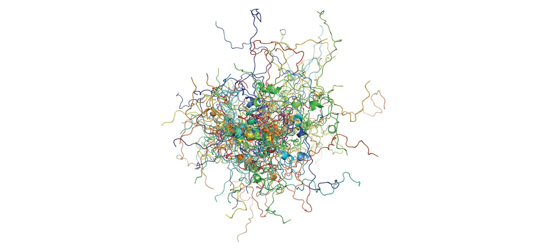
Many snapshots of the cancer protein p53, superimposed on top of one another and color-coded, illustrate the complex motion patterns of this intrinsically disordered protein. Some of the transient structural elements appear as “helix-like structures”. © Dániel Szöllősi / MPI-NAT
