26.08.2020
Authors Menezes R, Foito A, Jardim C, Costa I, Garcia G, Rosado-Ramos R, Freitag S, Alexander CJ, Outeiro TF, Stewart D, Santos CN Journal Antioxidants Citation Antioxidants (Basel). 2020 Aug 26;9(9):E789. Abstract Plants are a reservoir of high-value molecules with underexplored biomedical applications. With the aim of identifying novel health-promoting
24.08.2020
Emission States Variation of Single Graphene Quantum Dots
Authors Ghosh S, Oleksiievets N, Enderlein J, Chizhik AI Journal Journal of Physical Chemistry Letters Citation J. Phys. Chem. Lett. 2020, 11, XXX, 7356–7362. Abstract Graphene quantum dots (GQDs) are nanoparticles that consist of a nanometer-sized core of graphene with diverse chemical groups on its boundary. Due to their advantageous
21.08.2020
Neue Erkenntnisse über Lungengewebe bei Covid-19
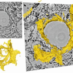
Physiker der Universität Göttingen und des Exzellenzclusters „Multiscale Bioimaging“ (MBExC) haben gemeinsam mit Pathologen und Lungenspezialisten der Medizinischen Hochschule Hannover ein Bildgebungsverfahren entwickelt, das eine hochauflösende und dreidimensionale Darstellung des geschädigten Lungengewebes nach schwerer Covid-19-Erkrankung ermöglicht. Mit einer speziellen Röntgenmikroskopietechnik konnten sie die durch das Coronavirus verursachten Veränderungen in der
21.08.2020
New insights into lung tissue in Covid-19 disease
Researchers led by Göttingen University develop new three-dimensional imaging technique to visualize tissue damage in severe Covid-19 (pug) Physicists at the University of Göttingen and the Cluster of Excellence “Multiscale Bioimaging” (MBExC), together with pathologists and lung specialists at the Medical University of Hannover, have developed a three-dimensional imaging technique
20.08.2020
Rosalind Franklin and the Advent of Molecular Biology
Authors Cramer P Journal Cell Citation Cell. 2020 Aug 20;182(4):787-789. Abstract Rosalind Franklin provided the key data for deriving the double helix structure of DNA. The English chemist also pioneered structural studies of colloids, viruses, and RNA. To celebrate the 100th anniversary of Franklin’s birth, I summarize her work, which
20.08.2020
3d Virtual Pathohistology of Lung Tissue from Covid-19 Patients based on Phase Contrast X-ray Tomography
Authors Eckermann M, Frohn J, Reichardt M, Osterhoff M, Sprung M, Westermeier F, Tzankov A, Werlein C, Kuehnel M, Jonigk D, Salditt T Journal Elife Citation eLife 2020;9:e60408. Abstract We present a three-dimensional (3D) approach for virtual histology and histopathology based on multi-scale phase contrast x-ray tomography, and use this
18.08.2020
3D virtual histology of human pancreatic tissue by multiscale phase-contrast X-ray tomography
Authors Frohn J, Pinkert-Leetsch D, Missbach-Güntner J, Reichardt M, Osterhoff M, Alves F, Salditt T Journal Journal of Synchrotron Radiation Citation J. Synchrotron Rad. (2020). 27, 1707-1719. Abstract A multiscale three-dimensional (3D) virtual histology approach is presented, based on two configurations of propagation phase-contrast X-ray tomography, which have been implemented
14.08.2020
Conformational dynamics of a G protein-coupled receptor helix 8 in lipid membranes
Authors Dijkman PM, Muñoz-García JC, Lavington SR, Kumagai PS, Dos Reis RI, Yin D, Stansfeld PJ, Costa-Filho AJ, Watts A Journal Science Advances Citation Sci Adv. 2020 Aug 14;6(33):eaav8207. Abstract G protein-coupled receptors (GPCRs) are the largest and pharmaceutically most important class of membrane proteins encoded in the human genome,
11.08.2020
Multicolor 3D MINFLUX nanoscopy of mitochondrial MICOS proteins
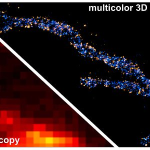
Authors Pape JK, Stephan T, Balzarotti F, Büchner R, Lange F, Riedel D, Jakobs S, Hell SW Journal Proceedings of the National Academy of Sciences Citation PNAS, Aug 2020, 202009364. Abstract The mitochondrial contact site and cristae organizing system (MICOS) is a multisubunit protein complex that is essential for the
11.08.2020
Proteine ganz nah
In einer ersten Anwendung der leistungsstarken MINFLUX-Nanoskopietechnik in der Zellbiologie haben Forscher um Stefan Hell und Stefan Jakobs die Verteilung einzelner Proteine in einem etwa 20 Nanometer großen Proteinkomplex innerhalb eines Zellorganells mehrfarbig und dreidimensional sichtbar gemacht. Diese Technik ist damit hundertmal schärfer als die herkömmliche Fluoreszenz-Lichtmikroskopie. Die MINFLUX-Nanoskopie erweist


