Researchers at the University Medical Center Göttingen (UMG) and the Max Planck Institute (MPI) for Multidisciplinary Sciences have shown, for the first time, how the genetic material of the Nipah virus replicates in infected cells. The virus can cause fatal encephalitis in humans. Using cryo-electron microscopy, the team led by MBExC member Hauke Hillen was able to visualize the three-dimensional structure of the viral “copying machine”. These findings could contribute to the future development of antiviral drugs for the treatment of Nipah virus infections. The results of the study have now been published in the journal Nature Communications.
“This is an important milestone because until now it was not known exactly what the RNA polymerase of the Nipah virus looks like and how it interacts with the viral RNA. Our data show that it is similar to the RNA polymerases of other related RNA viruses, such as Ebola, but has some special features,” says Hillen. The data also reveals how such a viral RNA polymerase uses the genomic viral RNA as a template for the copying process, and how it binds the newly produced product RNA and nucleotide building blocks.
Link to the press release (in English)
Link to the press release (in German)
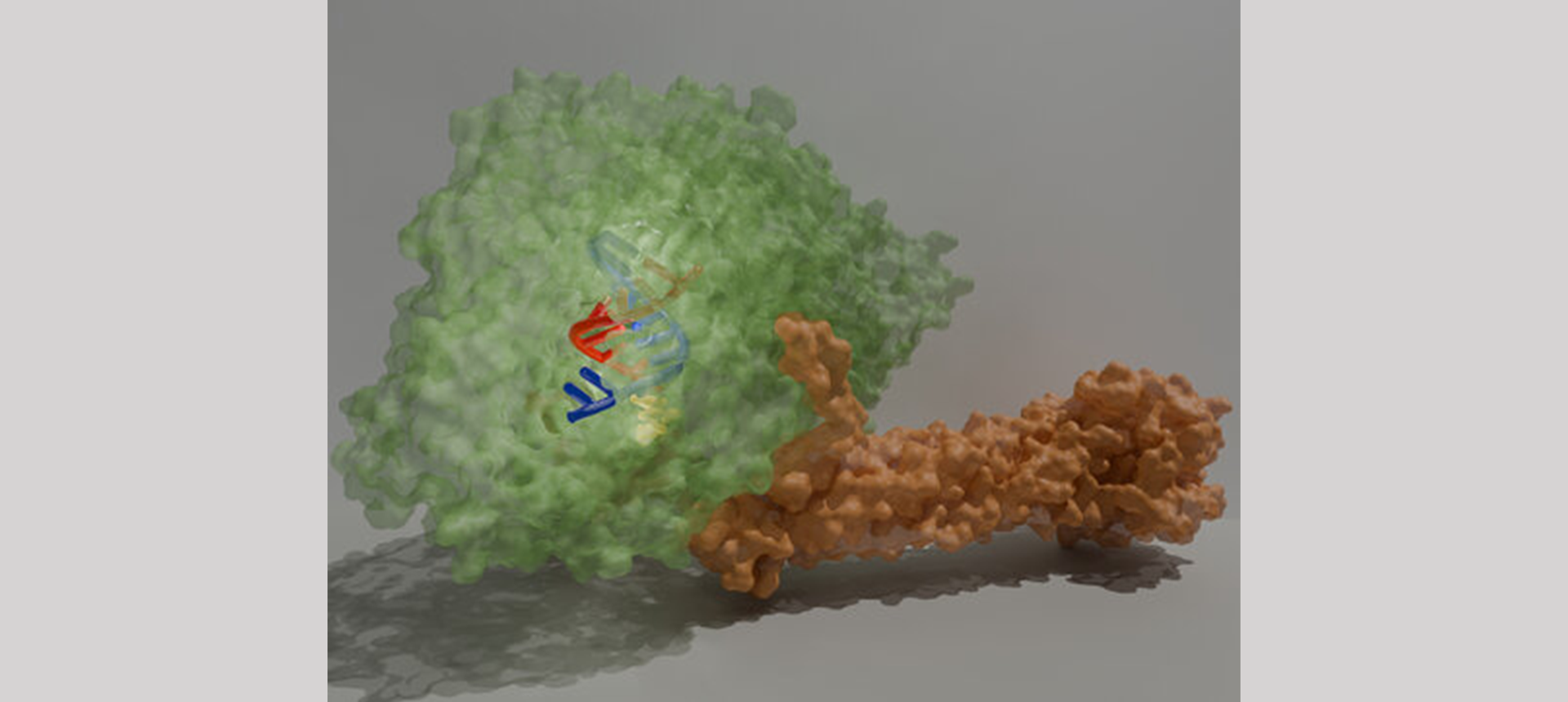
Artistic depiction of the 3D structure of Nipah virus RNA polymerase in the active state. The structure of the Nipah virus RNA polymerase is shown as a transparent surface representation (L protein in green, P protein in orange). The viral RNA, which serves as a template for the RNA polymerase, is shown in blue, the newly produced product RNA in red. The nucleotide substrate is shown in yellow. © Fernanda Sala / UMG