21

Home >> 2020 >> August >> 21

Sections through the three-dimensional reconstruction volume (upper left, grey) around a pulmonary alveolus with hyaline membrane (lower left, yellow). On the right, the images are superimposed. In the centre is the air bubble (alveolus). The electron density is represented by different shades of grey. On the inside of the air bubble is a layer of proteins and dead cell residues, the "hyaline membrane". This deposit, which can be represented in its three-dimensional structure for the first time by the new method, reduces the gas exchange and leads to respiratory distress. Photo: Tim Salditt, Marina Eckermann

21.08.2020

Neue Erkenntnisse über Lungengewebe bei Covid-19

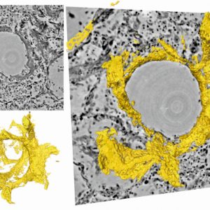
Physiker der Universität Göttingen und des Exzellenzclusters „Multiscale Bioimaging“ (MBExC) haben gemeinsam mit Pathologen und Lungenspezialisten der Medizinischen Hochschule Hannover ein Bildgebungsverfahren entwickelt, das eine hochauflösende und dreidimensionale Darstellung des geschädigten Lungengewebes nach schwerer Covid-19-Erkrankung ermöglicht. Mit einer speziellen Röntgenmikroskopietechnik konnten sie die durch das Coronavirus verursachten Veränderungen in der
Learn More
X

Open Positions

EN DE
X
X